
Laboratory Investigations in the Diagnosis and Management of Multiple Myeloma
Introduction
Multiple myeloma (MM) is a clonal plasma cell malignancy characterized by uncontrolled proliferation of plasma cells within the bone marrow and excessive production of monoclonal immunoglobulins. Laboratory investigations play a central role in diagnosis, staging, prognosis, treatment monitoring, and detection of relapse.
Advances in laboratory medicine, including serum free light chain assays, multiparametric flow cytometry, fluorescence in situ hybridization (FISH), and minimal residual disease (MRD) assessment, have transformed the management of multiple myeloma and improved patient outcomes.
Abstract
Multiple myeloma is a hematological malignancy characterized by clonal proliferation of plasma cells and production of monoclonal proteins. Laboratory investigations remain the cornerstone of diagnosis and management. Conventional investigations such as complete blood count, serum protein electrophoresis, immunofixation, and bone marrow examination continue to provide essential diagnostic information. Advanced techniques including flow cytometry, cytogenetics, FISH, next-generation sequencing, and MRD assessment enhance diagnostic accuracy and prognostic evaluation. This review discusses the role of laboratory investigations in the diagnosis, risk stratification, treatment monitoring, and relapse detection of multiple myeloma.
Role of Laboratory Investigations in Multiple Myeloma
Laboratory investigations are essential for:
Initial diagnosis
Disease staging
Risk stratification
Monitoring treatment response
Detection of minimal residual disease
Identification of relapse
Hematological Investigations
Complete Blood Count (CBC)
Common findings include:
Normocytic normochromic anemia
Leukopenia in advanced disease
Thrombocytopenia due to marrow infiltration
Clinical Significance
Anemia is one of the CRAB criteria used in diagnosing symptomatic multiple myeloma.
Peripheral Blood Smear
Characteristic findings:
Rouleaux formation
Anisocytosis
Poikilocytosis
Occasional circulating plasma cells
Erythrocyte Sedimentation Rate (ESR)
ESR is usually markedly elevated and often exceeds 100 mm/hour due to increased monoclonal immunoglobulins.
Biochemical Investigations
Serum Protein Studies
Typical findings include:
Elevated total protein
Reduced albumin
Increased globulin fraction
Serum Protein Electrophoresis (SPEP)
Purpose
Detection of M-protein
Quantification of disease burden
Monitoring treatment response
Characteristic Finding
A monoclonal spike (M-spike) in the gamma region.
Immunofixation Electrophoresis (IFE)
Used to:
Confirm monoclonal gammopathy
Determine immunoglobulin subtype
Identify light-chain restriction
Serum Free Light Chain Assay
Applications
Light-chain myeloma diagnosis
Monitoring disease progression
Early relapse detection
Important Biomarker
An involved/uninvolved free light chain ratio ≥100 is considered a myeloma-defining event.
Urine Investigations
Urine Protein Electrophoresis (UPEP)
Used for:
Detection of urinary monoclonal proteins
Quantification of Bence Jones proteinuria
Urine Immunofixation
Provides greater sensitivity than routine urine electrophoresis and confirms monoclonal light chains.
Assessment of Organ Damage
Renal Function Tests
Investigations include:
Serum creatinine
Blood urea nitrogen (BUN)
Estimated glomerular filtration rate (eGFR)
Serum Calcium
Hypercalcemia occurs due to increased bone resorption and is a major CRAB criterion.
Lactate Dehydrogenase (LDH)
Elevated LDH indicates:
High tumor burden
Aggressive disease biology
Poor prognosis
Beta-2 Microglobulin
An important prognostic marker incorporated into modern staging systems.
Bone Marrow Examination
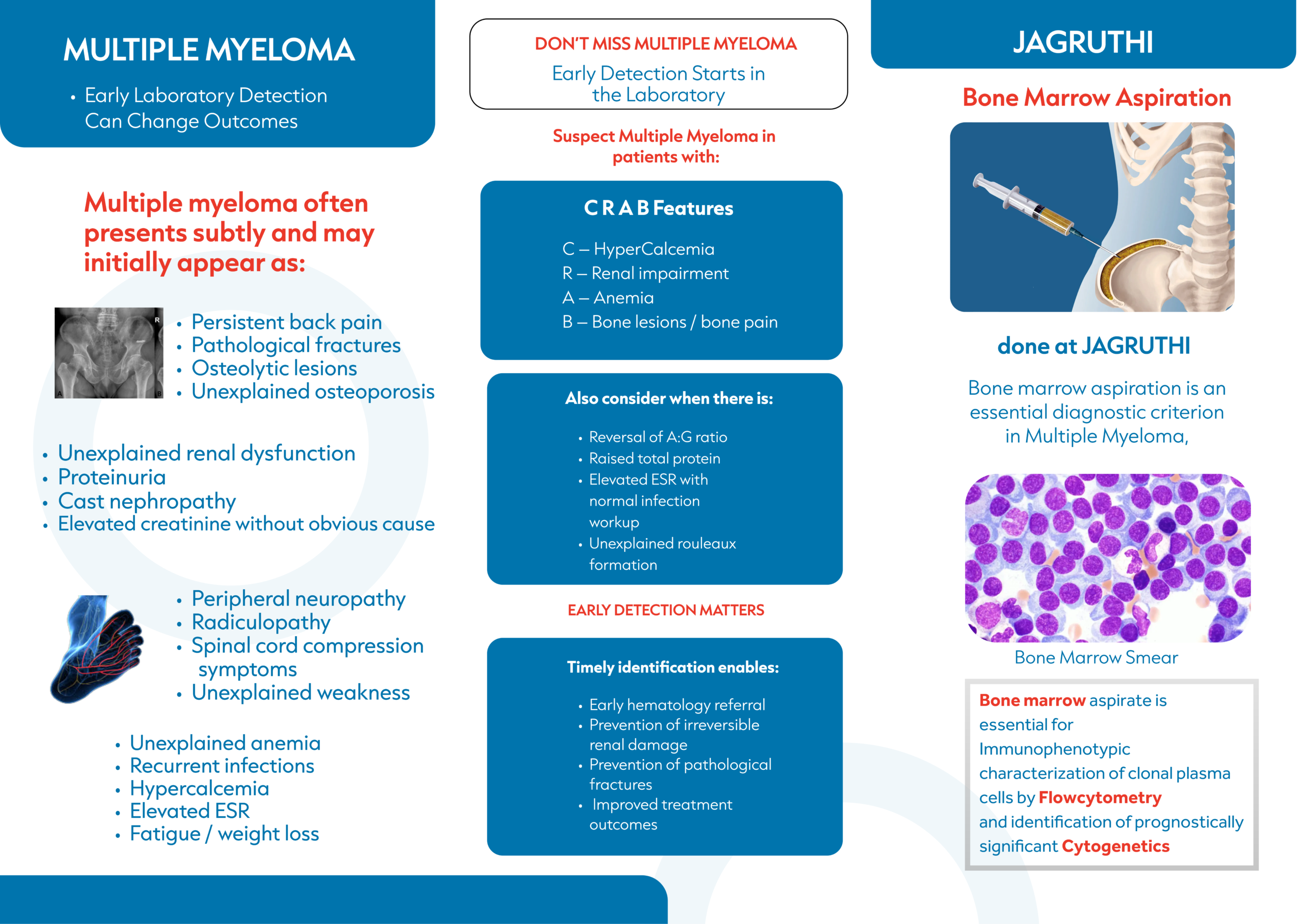
Bone Marrow Aspiration
Typical findings include:
Clonal plasma cells ≥10%
Abnormal plasma cell morphology
Bone Marrow Biopsy
Provides information regarding:
Marrow architecture
Plasma cell burden
Focal infiltration patterns
Flow Cytometry and Immunophenotyping
Typical Plasma Cell Markers
Positive markers:
CD38
CD138
CD56
Negative or reduced markers:
CD19
CD45
Clinical Applications
Diagnostic confirmation
Risk assessment
Minimal residual disease evaluation
Cytogenetic and Molecular Investigations
Fluorescence In Situ Hybridization (FISH)
High-risk abnormalities include:
del(17p)
t(4;14)
t(14;16)
Gain 1q
Next-Generation Sequencing (NGS)
NGS provides:
Comprehensive genomic profiling
Mutation detection
Precision medicine opportunities
Minimal Residual Disease (MRD) Assessment
Methods
Next-Generation Flow Cytometry
Sensitivity up to 10⁻⁵–10⁻⁶.
Next-Generation Sequencing
Sensitivity up to 10⁻⁶.
Clinical Importance
MRD negativity is associated with:
Improved progression-free survival
Better overall survival
Enhanced treatment outcomes
Monitoring Treatment Response
Complete Response (CR)
Negative serum immunofixation
Negative urine immunofixation
Less than 5% marrow plasma cells
Very Good Partial Response (VGPR)
More than 90% reduction in M-protein
Partial Response (PR)
At least 50% reduction in serum M-protein
Detection of Relapse
Laboratory indicators include:
Rising M-protein levels
Increasing free light chain concentrations
Reappearance of Bence Jones proteinuria
Increased marrow plasma cells
New CRAB features
Emerging Laboratory Technologies
Mass Spectrometry
Offers superior sensitivity for detecting residual monoclonal proteins.
Circulating Tumor DNA
Potential future applications include:
Molecular monitoring
Early relapse detection
Personalized treatment guidance
Artificial Intelligence
Emerging applications include:
Electrophoresis interpretation
MRD analysis
Prognostic modeling
Conclusion
Laboratory investigations remain fundamental to the diagnosis and management of multiple myeloma. Conventional diagnostic methods continue to provide essential information, while advanced technologies such as flow cytometry, FISH, next-generation sequencing, and MRD assessment have significantly enhanced diagnostic precision and prognostic evaluation. The integration of these laboratory modalities enables personalized treatment strategies and improved patient outcomes.
References
Rajkumar SV. Multiple Myeloma: Recent Advances in Diagnosis and Management.
International Myeloma Working Group Diagnostic Criteria.
NCCN Clinical Practice Guidelines for Multiple Myeloma.
Palumbo A, Anderson KC. Multiple Myeloma. New England Journal of Medicine.
Kumar S, Paiva B, Anderson KC, et al. MRD Assessment in Multiple Myeloma.